Regular screening mammography is recommended from the age of 40 for all women with an average risk of breast cancer. For women who have a family history of breast cancer (mother, grandmother, sister), mammograms are usually advised from the age of 35.
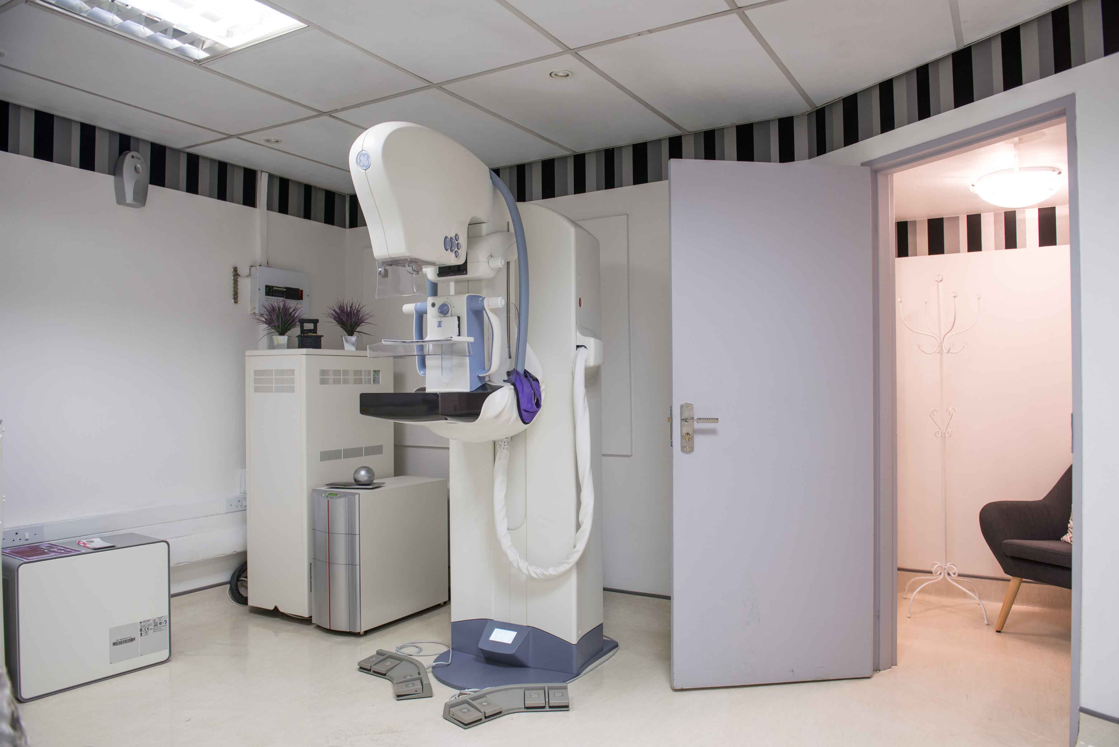
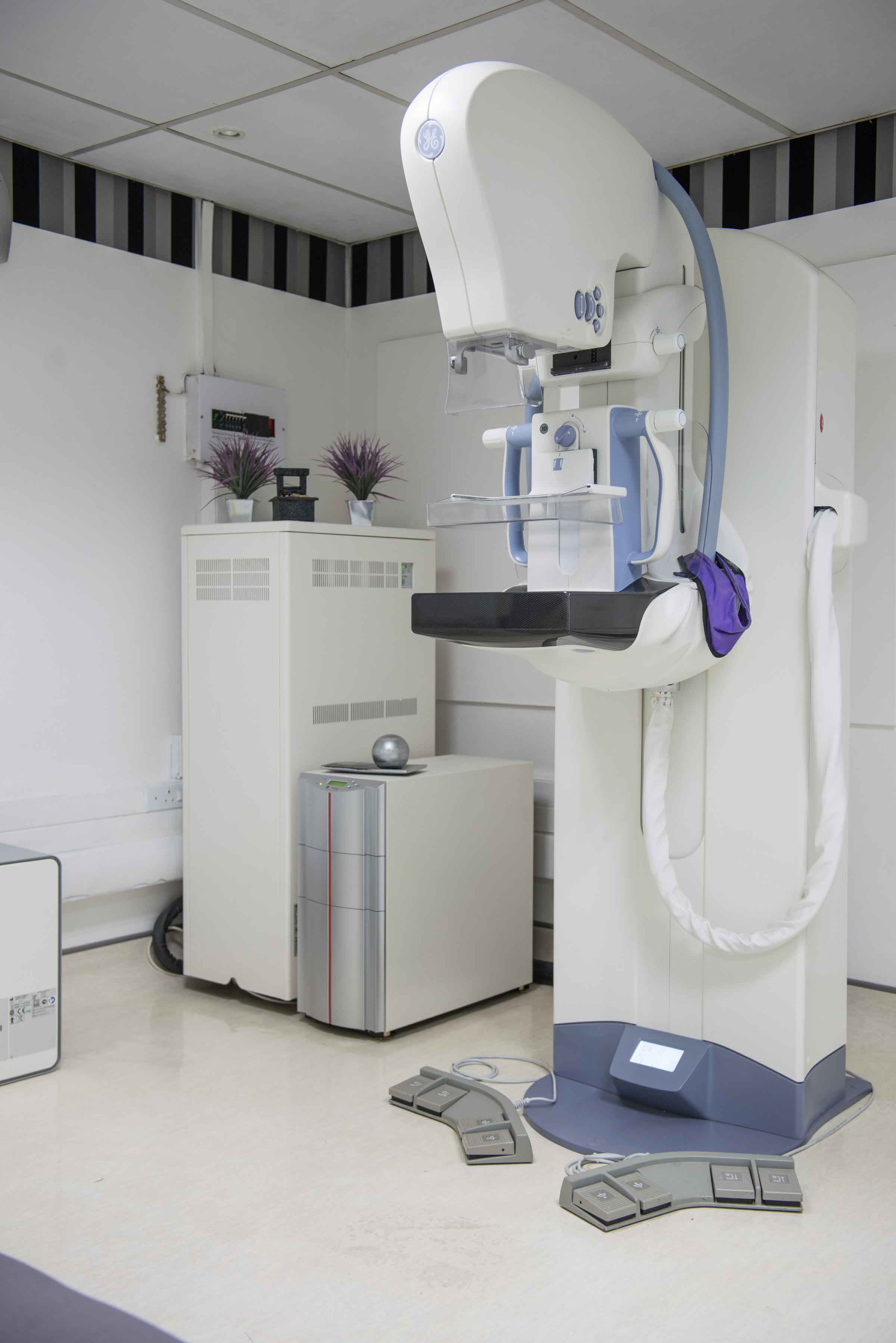
Mammography involves the use of low-dose x-rays to obtain non-invasive, high contrast/high resolution detailed imaging of the breasts. These images can help detect early stages of breast cancer, demonstrating changes in the breasts long before they can be felt clinically. Recent advances in imaging include digital mammography, contrast-enhanced mammography, breast tomosynthesis and breast MRI, all available at NRS.
General Information
A complete radiographic breast evaluation includes clinical history, breast palpation, a mammogram and a breast ultrasound. Mammography and breast ultrasound are both required as they complement each other. All mammograms are performed by qualified mammography radiographers. The findings are then reviewed by an onsite diagnostic radiologist and if necessary, additional tests or intervention such as aspiration or biopsy will be recommended to determine benign or malignant disease.
Mammographic interpretation can be difficult because the normal breast tissue appears different for each patient and the imaging appearance can be altered if there has been breast surgery. Comparison with any earlier examinations can assist to detect new subtle changes. Where possible, always provide the interpreting radiologist with your previous breast imaging examinations.
It is very important to realize that not all breast cancers can be seen on mammography. Interpretation is particularly limited when breast tissue is increased in density or in the presence of implants, which can obscure tissue behind them because they are not transparent at x-ray imaging.
NRS offers digital mammography which allows for a lower radiation dose. Images are available for review on a computer and can be stored digitally, printed and copied to CD. Breast Tomosynthesis (available at Ahmed Kathrada Hospital) is an advanced form of imaging which acquires a set of multiple images of each breast and allows for improved lesion detection and characterisation, particularly within dense breast tissue. Contrast-enhanced mammography is an adjunct to this, allowing for further lesion characterisation. Breast MRI is available at Waterfall City Hospital.
Although mammography involves radiation, the benefits of an early diagnosis, far outweigh the risks. The lowest possible radiation dose is used to obtain the study. Radiation doses are carefully monitored at all our sites.
Always confirm aspiration or biopsy results with your doctor and comply with the recommended follow-up interval. Regular self-examination of the breasts is important.
How the Test is performed
Once you have provided the relevant clinical information, the radiographer will position you standing in front of the mammogram machine and place one of your breasts between two plastic plates. These plates gradually compress your breast so that radiation is reduced and the image produced demonstrates the most detail. Each compression should only last a few seconds, causing discomfort rather than pain. Because most breast cancers develop from glandular tissue and occur centrally and laterally in the breast, it is very important that these areas show up very clearly on the film. Usually, two views of each breast in different positions are acquired. Additional views may be necessary in certain circumstances to further evaluate an area of concern.
Your co-operation with the radiographer is essential to obtain the best images. Once the radiographer is satisfied with the quality of the films, you will proceed to have an ultrasound of your breasts. Please allow sufficient time for the Radiologist to examine your images professionally.
Once reviewed by a radiologist, suspicious abnormalities may need additional tests such as aspiration or biopsy for a definitive diagnosis.
Preparing for the Test
Avoid wearing deodorant, powder or lotion under your arms or on your breasts as these substances may create artifacts on the images and obscure image detail.
Ensure that previous mammogram studies are brought with you on the day of your examination for the radiologists to view.
As your breasts are usually tender around menstruation, plan your examination around one week after menstruation to avoid further discomfort during breast compression.
A skirt or pants is preferable. Your top will need to be removed. A loose-fitting gown will be provided for you.
Inform the radiographer if you are pregnant or breast feeding.
If an interventional biopsy or aspiration is indicated, inform the radiologist of any blood thinning medication that you may be taking.
After the Test
There may be mild breast discomfort or tenderness after the study. Please inform the radiographer if there is significant pain.
Pain after biopsy or aspiration can be managed with cold packs and basic analgesia such as paracetamol. Aspirin should be avoided. There may be regional bruising after the procedure which is temporary. Please inform your doctor of there is excessive bleeding, redness, warmth or increasing tenderness.
Breast Biopsies
After your mammogram, the radiologist may suggest a breast biopsy to determine whether pathology on your mammogram is benign or malignant.
Several breast biopsy procedures are used to obtain a tissue sample from the breast. Your radiologist may recommend a particular procedure based on the size, location and other characteristics of the breast abnormality.
For most of the biopsies, you will receive an injection to numb the area.
The different types of biopsies:
Ultrasound-guided Fine-needle aspiration biopsy (FNA)
This is the simplest type of breast biopsy and may be used to evaluate a lump that has been seen on your ultrasound or mammogram. For this procedure, you lie on a table in the ultrasound room. While steadying the lump with one hand, your radiologist uses the other hand to direct a very thin needle into the lump, using ultrasound to guide the needle. At least three samples are obtained. Fine-needle aspiration is a quick way to distinguish between a fluid-filled cyst and a solid mass. If the mass is solid, a tissue sample will be obtained.
Ultrasound-guided Core needle biopsy
Depending on the location of the mass, a variety of imaging techniques can be used to guide a core needle biopsy to obtain the tissue sample. These imaging may include a mammogram, ultrasound or MRI. Ultrasound guidance is used at our facilities.
This type of breast biopsy may be used to assess a breast lump that is visible on one of the above imaging techniques or that your doctor feels (palpates) during a clinical breast exam. The specialized radiologist uses a thin, hollow needle to remove tissue samples from the breast mass.
Several samples can be obtained and then analysed by the pathologist to identify features indicating the presence of disease.
Ultrasound-guided Vacuum-assisted biopsy
A special probe is inserted into the lesion. A vacuum and cutting device is then used to draw the tissue samples into the probe. This procedure is able to remove more tissue than a core needle biopsy and in some cases, small abnormalities may be completely removed.
Stereotactic biopsy
Not available at NRS.
This type of biopsy uses mammograms to pinpoint the exact location of suspicious pathology within your breast. It is necessary when a suspicious finding identified at mammography is not visible at ultrasound and can therefore only be accessed by using mammographic guidance. For this procedure, you generally are in the same position as for the mammogram or if possible you may have the procedure in a seated position. You may need to remain in this position for +/- 15 minutes. The radiographer will make you as comfortable as possible while the biopsy is being done.
Insertion of a Marker
Occasionally, your breast surgeon may request insertion of a small (2mm) marker into a breast cancer lesion. The marker is inserted under ultrasound guidance through a hollow needle. A plunger is used to push the marker into the desired area. Following placement of the marker, a single mammogram image is acquired to confirm correct placement.


Branches
Netcare N17 Hospital
bernita@nrsmail.co.za/ 011 815 2814
Ahmed Kathrada Private Hospital
aisha@nrsmail.co.za / 011 852 8820
Netcare Waterfall City Hospital
corne@nrsmail.co.za / 010 612 6669
/ 010 612 0612 / 011 304 7751
071 246 5759
Contact Head Office
National Radiology Services Inc.
12 Stirrup Lane
Woodmead
1682
Tel: 010 612 0271 / 010 612 0612