General X-Rays
Plain film radiography is the oldest form of imaging. This painless procedure uses the physical properties of the X-ray to create a 2D image which displays body tissues of different density and composition and aids clinicians in managing a variety of medical conditions.
General Information
Radiography is the imaging of internal body structures using X-rays, which are a form of radiation similar to visible light, radiowaves and microwaves.
X-radiation has a very high energy level that allows the X-ray beam to penetrate through the body and create an image or picture. The image is created due to the X-ray beam being absorbed differently by different structures or parts in the body. A dense structure like bone absorbs a high percentage of the X-ray beam, whilst low density structures like soft tissues absorb a small percentage.
How the Test is Performed
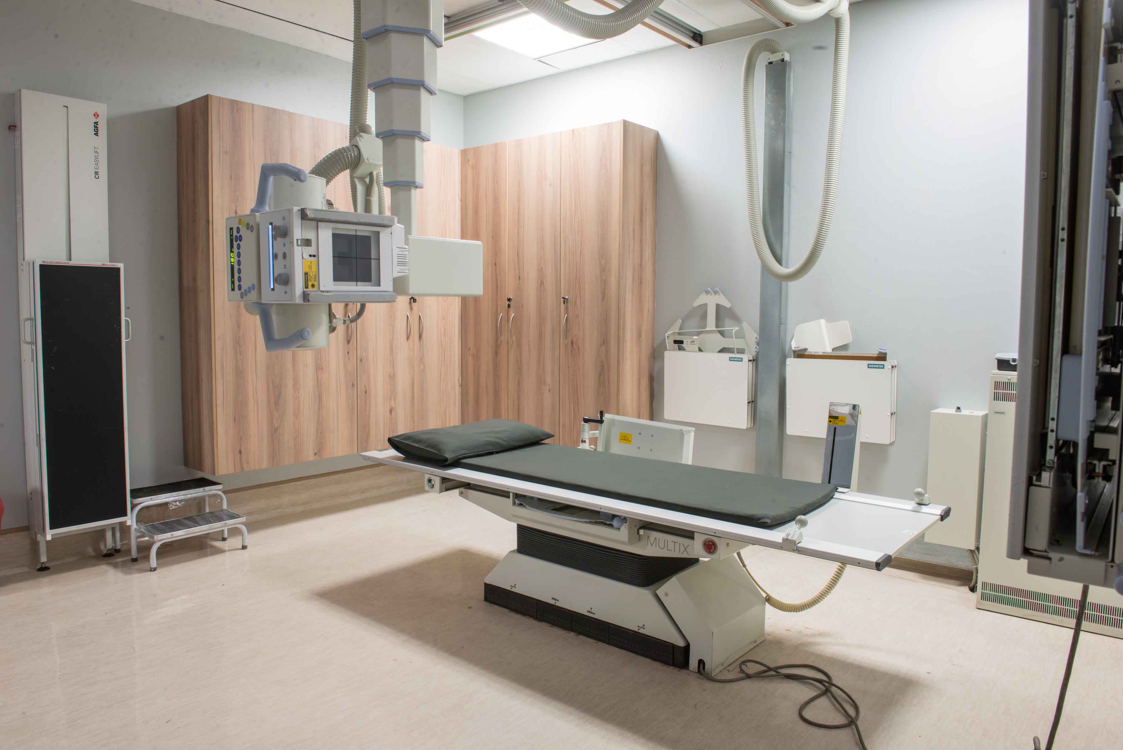
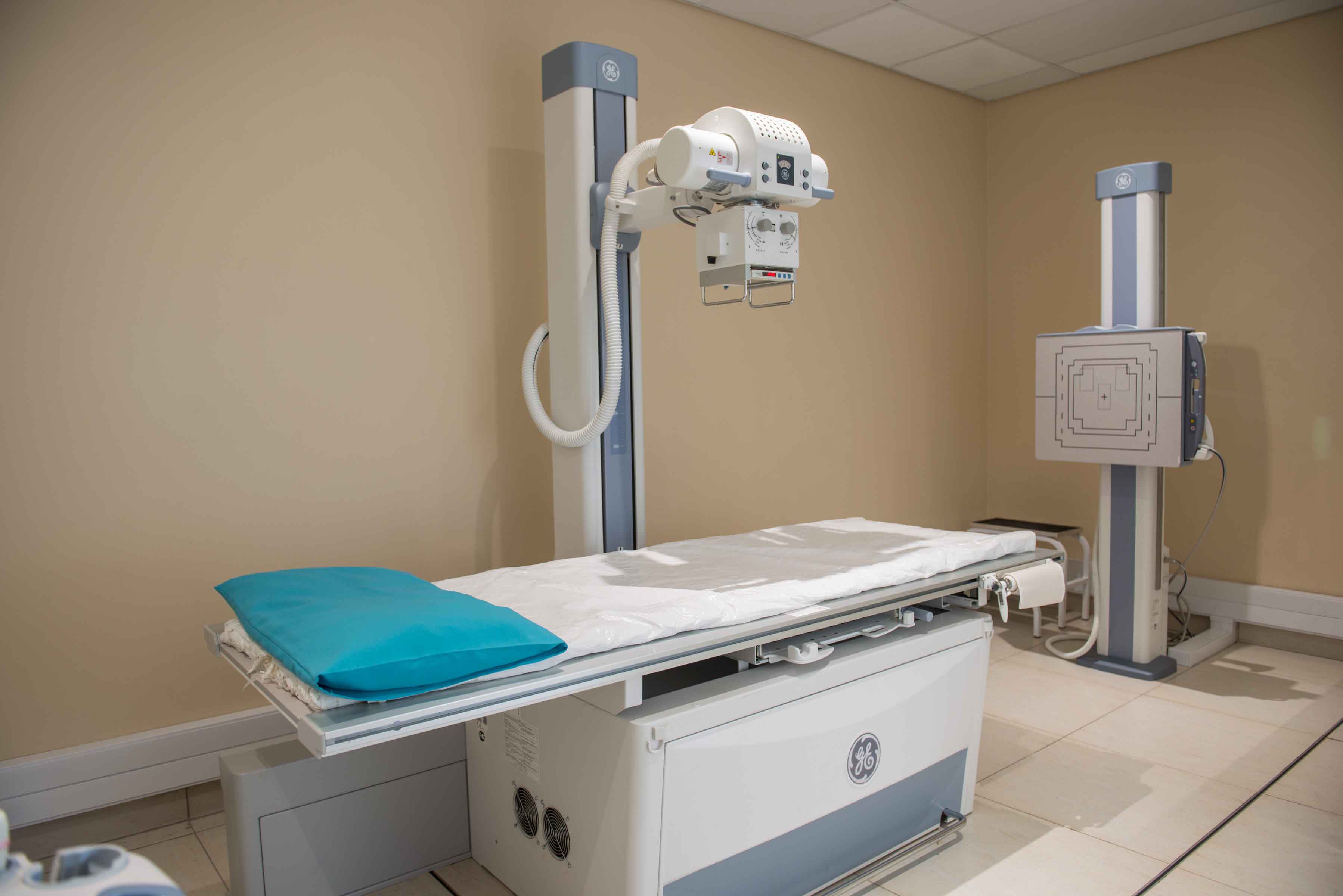
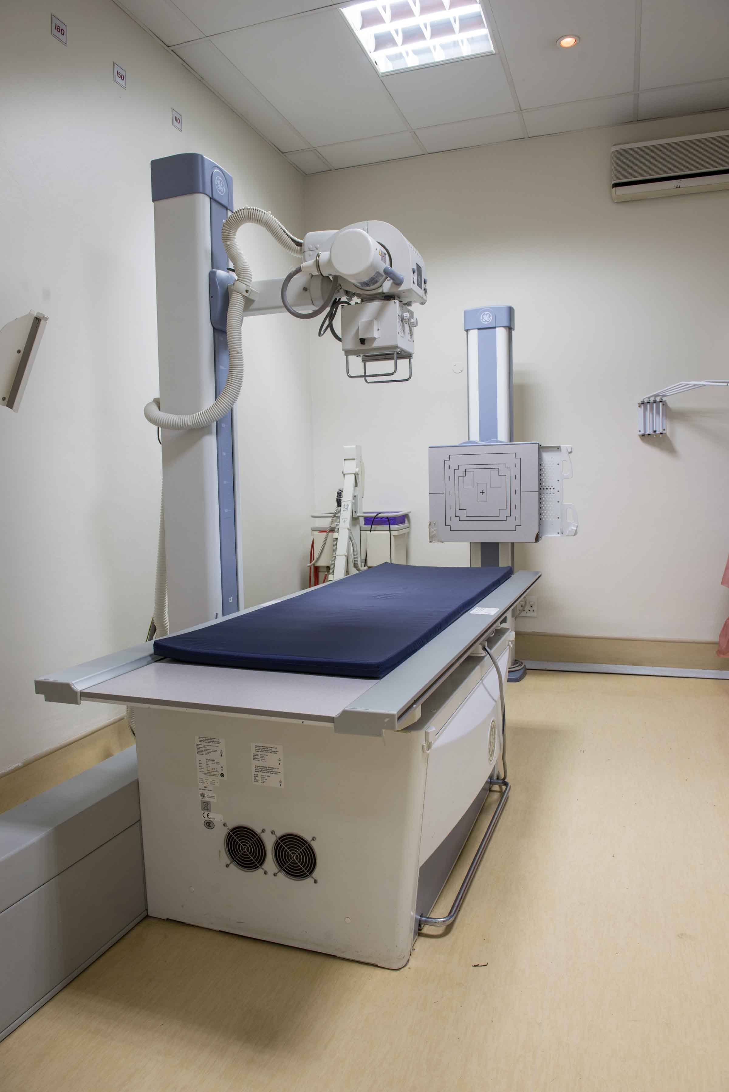
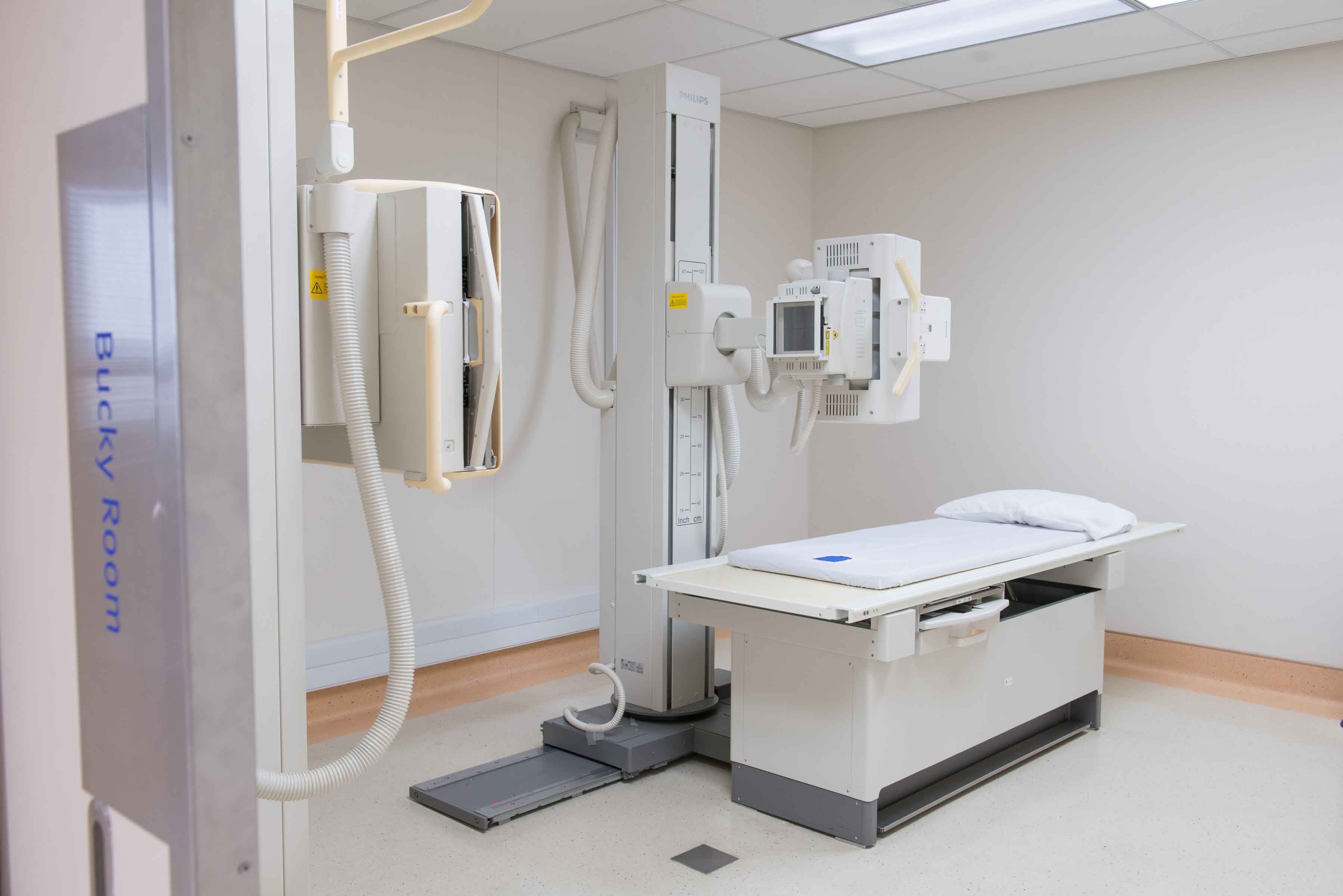
Radiographs (“x-ray films’) are produced by the transmission of x-rays through a patient to a capture device with a film which is then processed. The X-ray beam is a form of electromagnetic radiation. “Plain” films are the most frequent examinations in the radiology department. Examples of these are chest, spine, skull x-rays – in fact any area of the skeleton.
A radiographer performs this simple and painless procedure. Generally, more than one view is acquired. Depending on the region of interest, you may be required to stand, sit, or lie down. Chest and abdominal radiographs often require that you hold your breath while the exposure is made. Your co-operation with the radiographer will ensure that the best quality imaging is obtained. The images can be captured onto an x-ray film or copied onto a CD.
Lead protection may be used to avoid radiation exposure to areas of the body that are not being imaged. Special care is taken with paediatric patients. We are committed to obtaining quality images, whilst minimizing the risk of radiation exposure to patients from imaging procedures whenever and wherever possible.
While it only takes a few minutes to perform your examination, extra time is needed to process and review your images. All radiographs are reviewed by our radiologists. Please allow us sufficient time to do this professionally.
Preparation for the Test
No preparation is required for these examinations.
Inform the health care provider if you are pregnant. X-rays are generally avoided during the first three months of pregnancy. If the clinician decides that imaging is necessary during pregnancy, limited views will be acquired and appropriate shielding will be utilised.
You may be asked to wear a hospital gown and remove all jewellery.

Branches
Netcare N17 Hospital
bernita@nrsmail.co.za/ 011 815 2814
Ahmed Kathrada Private Hospital
aisha@nrsmail.co.za / 011 852 8820
Netcare Waterfall City Hospital
corne@nrsmail.co.za / 010 612 6669
/ 010 612 0612 / 011 304 7751
071 246 5759
Contact Head Office
National Radiology Services Inc.
12 Stirrup Lane
Woodmead
1682
Tel: 010 612 0271 / 010 612 0612